ORTHOPAEDICS
WITH ARTIFICIAL JOINTS FOR A BETTER QUALITY OF LIFE
Arthroplasty is the medical speciality for implanting artificial joints. These can be used in the shoulder, knee, hip, hand and fingers, among others, but in principle it is possible to replace almost any joint. In arthroplasty, specialised knowledge and in-depth expertise are essential in order to guarantee the best possible care for the client. The aim is to give you a life without pain and a significant improvement in joint function. Both of these ensure a significant increase in quality of life. And it is precisely this quality that we stand for at the ETHIANUM. To ensure that you can expect the best possible medical care and advice, we work together on an interdisciplinary basis in this area. In addition, the ETHIANUM Clinic has state-of-the-art medical equipment that can always be used and deployed in your favour and, in particular, without waiting times.
Arthroplasty is the last resort to enable you to lead a pain-free life. We have summarised the different areas and applications of arthroplasty for you.
ORTHOPAEDICS
OUR DOCTORS AT THE ETHIANUM
Get to know our specialists, who work hand in hand and with great commitment for you in the field of arthroplasty. Find out more about our experts in the field of arthroplasty.

FELIX ZEIFANG
–
Specialist in orthopaedics and trauma surgery
Prof Dr Felix Zeifang is a specialist in orthopaedics and trauma surgery. He specialises in shoulder, foot and elbow surgery and sports medicine. His individualised treatment concepts lead to a very high success rate.

GÜNTER GERMANN
–
Founder, Medical Director and Plastic Surgeon
Prof Dr Günter Germann is the founder and Medical Director of the ETHIANUM Clinic Heidelberg. The plastic surgeon can look back on an extremely successful career as a plastic and aesthetic surgeon. Clients greatly appreciate his talents as a hand surgery specialist and excellent microsurgeon.

JÖRG HOLSTEIN
–
Specialist in orthopaedics and trauma surgery
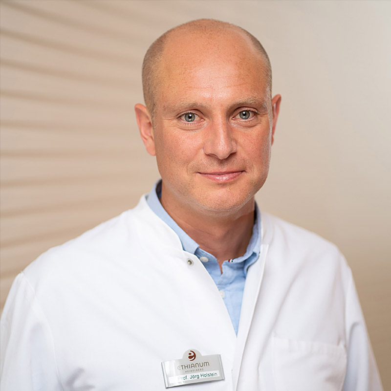
Prof. Dr. Jörg Holstein is a specialist in orthopaedics and trauma surgery. He specialises in hip and knee endoprosthetics. His minimally invasive and muscle-sparing surgical techniques and his vision for the benefit of his clients make him particularly popular.

PHILIPP A. MICHEL
–
Specialist in orthopedics and trauma surgery
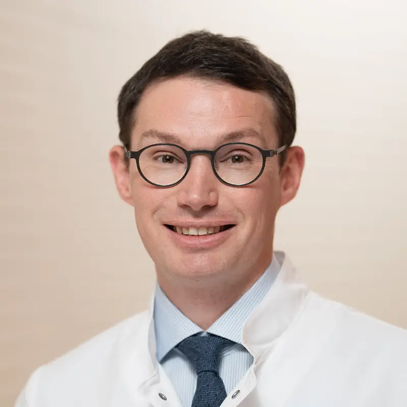
Priv.-Doz. Dr. Philipp A. Michel is a proven specialist in knee and hip arthroplasty with many years of clinical experience and excellent scientific qualifications. He will be joining the team of doctors at ETHIANUM in Heidelberg in July 2025.
Learn more about his robot-assisted surgical method.
ORTHOPAEDICS
SHOULDER JOINT ARTHROPLASTY
A great deal of experience is required when implanting a shoulder endoprosthesis. The surgeon must not only have an excellent command of the bony fixation of the endoprosthesis, but also of soft tissue management. This is because the correct positioning of the prosthesis has an influence on the service life of the prosthesis, regardless of whether it is an anatomical or inverse prosthesis. The aim is to position the new joint correctly with the right soft tissue tension.
Shoulder joint arthroplasty and possible surgical procedures
Shoulder joint arthroplasty is possible and conceivable for the following diagnoses:
- Osteoarthritis as a result of degenerative wear and tear
- Rheumatoid arthritis
- Fracture of the humeral head
- Necrosis of the humeral head
- Cufftear arthropathy (secondary arthrosis after major and chronic rotator cuff rupture)
The choice of prosthesis type depends on the degree of remaining shoulder function, the condition of the rotator cuff, the bone substance and the age of the patient.
- Anatomical total shoulder prostheses: These are generally implanted in patients with a still functional rotator cuff. The humeral head and glenoid cavity are replaced. Ceramic or pyrocarbon heads are now also used. The new materials are intended to extend the durability of the prostheses.
- Inverse shoulder prosthesis: The highest growth rates of all implanted joint prostheses in humans are recorded for the inverse shoulder prosthesis. It has shown very good results over the course of more than ten years. An inverse prosthesis should be implanted in patients with a defective rotator cuff, a fractured humeral head, non-functional or failed plate osteosynthesis and larger bone defects.
- Shoulder hemiprosthesis: A complete shoulder joint replacement in which both the head and socket are replaced is not always the ideal measure. Particularly in young patients, the choice of endoprosthesis should also be made in view of the fact that they will still have a long life. Surgeons can achieve good results with partial prostheses by replacing the humeral head. The socket must not yet show any significant signs of osteoarthritis. The less the glenoid is involved in the wear, the better. Caution: An unreplaced glenoid cavity can be problematic in the medium to long term, as it is still exposed to the degenerative process.
After the operation: The more mobile the muscles, tendons and ligaments are, the better the functional result of an arthroplasty reconstruction will be. Patients should therefore not wait too long if joint surfaces in the shoulder are already severely damaged.
The procedure is carried out as part of an inpatient hospital stay, which only lasts a few days. The arm is immobilised in a shoulder abduction cushion for a week, but the joint can be moved again from the very first day after the shoulder operation. Showering or washing hair is possible after about two days. Everyday activities such as eating with the operated arm should be possible again after three days at the latest.
The hospitalisation is followed by outpatient or inpatient rehabilitation. This is agreed with the patient in advance. Physiotherapy treatment is then continued on an outpatient basis. Here too, the quality of the physiotherapy follow-up treatment is decisive for the final result.
It is usually possible to resume everyday activities after six weeks; you should be able to resume more demanding sporting activities such as playing tennis or golf after three months at the latest.
ORTHOPAEDICS
HIP JOINT ARTHROPLASTY
If the advanced stage of hip joint osteoarthritis can no longer be treated with conservative measures, joint replacement surgery using the AMIS technique will be the specialist’s recommendation. Before an artificial hip joint is implanted, the correct position and size of the endoprosthesis is precisely planned on the X-ray image using special software. Osteoarthritis primarily destroys the cartilage and neighbouring bone of the femoral head and the acetabulum. Accordingly, these structures must be replaced by the artificial joint, which then takes over the function of the original joint.
Structure of a hip joint endoprosthesis and possible surgical procedures
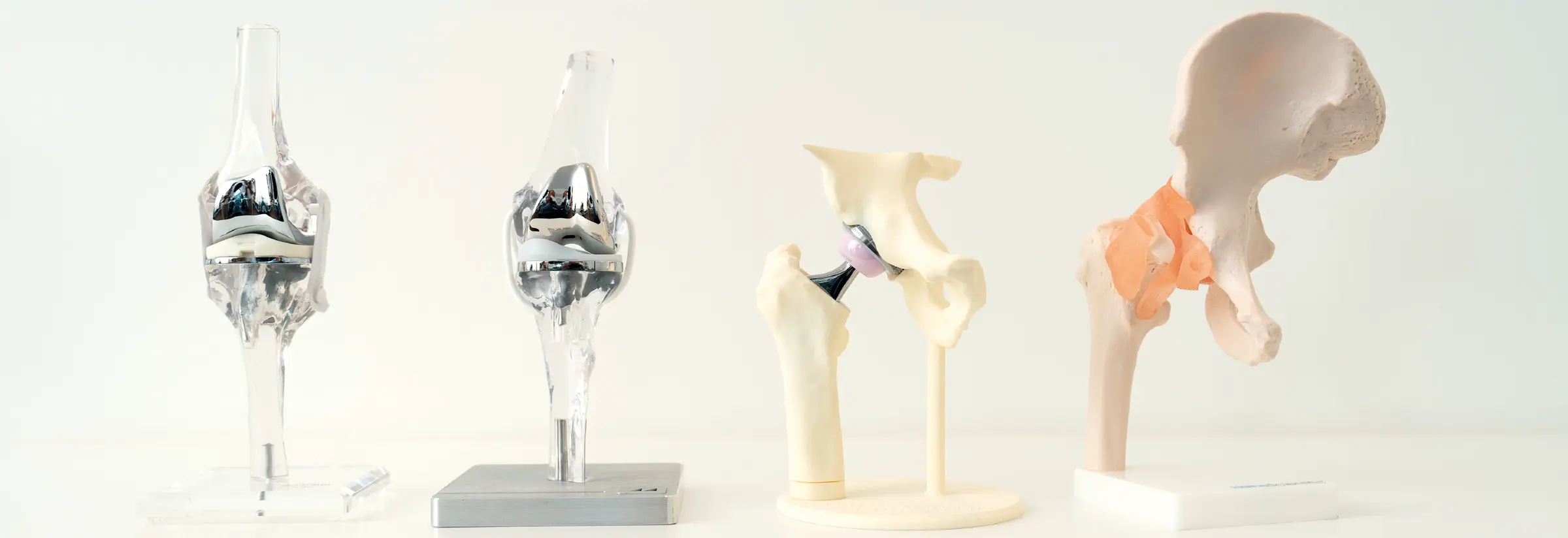
Endoprosthesis models in which an artificial ceramic femoral head is placed on a titanium stem that is clamped in the femur have become established. The artificial hip joint cup is also made of titanium and is implanted “press-fit” into the pelvic bone. An inlay made of ultra-highly cross-linked polyethylene or ceramic is inserted into the cup. Similar to cartilage, ceramic and ultra-highly cross-linked polyethylene have very good sliding properties so that there is hardly any friction between the new joint partners. To summarise, the artificial hip joint consists of four components:
- A titanium stem, which is anchored in the femur
- A ceramic head that is attached to the titanium hip stem
- A titanium cup that is anchored in the pelvic bone
- A ceramic or ultra-highly cross-linked polyethylene inlay that is inserted into the titanium cup
If the bone quality is poor, for example as a result of osteoporosis, the endoprosthesis stem or cup can be anchored in the femur or pelvic bone with bone cement as an alternative to the press-fit implantation technique. In this case, the stem is made of a cobalt-chrome alloy. The aim of both techniques is to anchor the endoprosthesis so firmly in the bone that you can walk and stand with your full body weight immediately after the operation.
The four approaches to the hip joint:
- The dorsal (posterior) approach
The dorsal approach leads through the gluteal muscles and through a muscle group that turns the leg outwards (“external rotators”) from behind to the hip joint. This approach provides quick access to the hip and a good overview during the operation. Unfortunately, the gluteal muscles and the external rotators are damaged during the approach. This can lead to muscular weakness and thus delay or impair rehabilitation after the operation. There is also an increased risk of joint dislocation, i.e. dislocation of the artificial femoral head popping out of the acetabulum later on with this approach. - The lateral approach
This approach leads through the lateral hip stabiliser muscles (“abductors”) to the hip joint. Similar to the posterior approach, the lateral approach provides quick access to the hip and gives the surgeon a good view of the joint. However, the lateral approach also damages a very important muscle group, namely the abductor group. This means that the lateral approach to the hip has similar disadvantages to the posterior approach. Muscular weakness delays rehabilitation and can favour joint dislocation. - The anterolateral approach
This approach does not pass through the muscles, but between muscles to the hip joint. The anterolateral approach is therefore generally gentler than the posterior and lateral approaches.
However, in order to obtain a good overview of the hip joint, it is necessary to hold the important lateral hip stabiliser muscle (gluteus medius muscle) to the side with hooks. This often causes injury to the muscle, which in turn impairs rehabilitation after the operation. Studies have also shown that the hooks often injure the nerve that innervates the so-called sprinter muscle (tensor fasciae latae muscle), which in turn can lead to partial leg weakness after the operation. - The anterior approach
Similar to the anterolateral approach, the anterior approach does not lead through the musculature, but between different muscles to the hip joint. One special feature: no nerves cross the approach. Studies have shown that the anterior approach is associated with the lowest risk of muscle damage and joint dislocation. This makes the anterior approach a particularly gentle approach technique.
The minimally invasive AMIS technique (DAA)
In the context of an artificial hip joint, minimally invasive surgical technique means not only the shortest possible skin incisions, but above all the protection of important functional body structures, in particular the muscles and tendons. The AMIS technique represents a consistent further development of the anterior approach. Recent studies have shown that patients operated on using the AMIS technique suffer less blood loss, can leave hospital earlier, are mobile more quickly and suffer fewer complications such as joint dislocations. You can find out more about the AMIS technique here.
ORTHOPAEDICS
KNEE JOINT ARTHROPLASTY
The right time for an artificial knee joint, a knee joint endoprosthesis, cannot be determined solely on the basis of an X-ray or MRI image – the restrictions that a person experiences in their daily professional and private life are the decisive factor. If conservative therapies no longer provide any relief, if the level of suffering and the restrictions on your quality of life caused by the symptoms of osteoarthritis are so great that the current condition is no longer acceptable to you, then the implantation of a knee joint endoprosthesis may make sense.
At the ETHIANUM, our specialists have mastered both the latest robot-assisted procedures and proven conventional techniques in order to implement the right solution for you with the utmost precision.
Knee joint endoprosthetics and possible surgical procedures
The advantages of robot assistance with CORI™
The ETHIANUM is one of the few clinics in the region to use the innovative CORI™ robotic system from Smith & Nephew. This technology significantly supports our experienced surgeons in optimally adapting the joint replacement to your individual anatomy and performing the procedure with the utmost precision.
How does the CORI™ system work?
- Image-free 3D planning: The system creates a precise 3D model of your knee in real time during the operation, without the need for a prior CT scan.
- Intelligent instrument guidance: The surgeon guides the instrument, the robotically controlled Reimer helps to implement the precise planning exactly and protects against unwanted deviations during bone processing. The surgeon retains complete control at all times.
- Dynamic ligament management: The system enables precise assessment and adjustment of the ligament tension over the entire range of motion – a decisive factor for the stability and function of the prosthesis.
Possible advantages of robotic assistance for you as a patient: - Maximum accuracy & customization: Studies show that robot-assisted systems can enable greater accuracy in the positioning and alignment of implants. The implant is adapted precisely to your anatomy. This is an important prerequisite for longevity, optimized function and a more natural feeling of movement.
- Potentially faster recovery & tissue sparing: The high precision can help to spare the surrounding healthy tissue and bone. Research results indicate less blood loss, less post-operative pain and in some cases shorter hospital stays compared to traditional procedures.
There are basically two types of knee endoprostheses:
- Partial joint replacement (sled prosthesis): However, if the osteoarthritis is only limited to one part (compartment) of your knee joint and the important cruciate and collateral ligaments are intact, a partial prosthesis (unicompartmental endoprosthesis or sled prosthesis) can be an excellent, gentler alternative to a total joint replacement.
- Advantages of the sled prosthesis:
- Preservation of healthy structures: Only the damaged part of the joint is replaced. Healthy bone areas, cartilage and important ligaments are preserved. This is the decisive advantage over a full prosthesis.
- More natural kinematics: As the patient’s own joint mechanics are largely retained, patients often report a more natural knee feeling and better mobility after a sled prosthesis.
- Minimally invasive: The procedure is generally smaller and gentler on the tissue.
- Faster rehabilitation: Recovery is often faster after a sled prosthesis. Robotics for the sled prosthesis: The CORI™ system really comes into its own with the sled prosthesis, where exact positioning is particularly critical for long-term success and healthy structures should be spared as much as possible. The unsurpassed precision of the robotic assistance helps to optimally position and adjust the partial prosthesis. Some studies suggest that patients can experience faster functional improvement and greater satisfaction in the first few months after a robot-assisted sled prosthesis.
- Total knee arthroplasty (TEP): If several parts (compartments) of your knee joint are severely damaged by osteoarthritis or if certain ligament instabilities are present, the complete replacement of the joint surfaces (total knee arthroplasty, knee TEP) is the appropriate solution to comprehensively eliminate pain and restore the function of the entire joint. Robotics for knee TEP: The advantages of CORI™ robotic assistance described above are particularly valuable for total knee arthroplasty. The precise alignment of the components and the exact management of the ligament tension not only enable optimal function and stability of the joint replacement. They also allow the surgeon to precisely implement modern alignment concepts such as kinematic alignment . While traditional methods often aim for a purely mechanical, straight alignment, kinematic alignment aims to reconstruct the patient’s original, individual joint line and axis of movement before osteoarthritis as best as possible. Every person has a slightly different knee anatomy and movement; the kinematic alignment takes this into account. The aim is to create a knee joint that feels as “normal” and natural as possible for the patient and enables harmonious interaction with the body’s own ligaments and muscles. The CORI™ system provides ideal support for the surgeon.
- Alternative: Proven conventional surgical technique – the medial pivot concept
Of course, robot-assisted surgery is not the only option. We also offer excellent solutions for patients who prefer surgery using proven conventional techniques or for whom there are specific reasons for not using robotics. Prof. Holstein, for example, successfully uses the medial pivot concept for conventional total knee arthroplasty.
What is the medial pivot concept?
These special prosthesis designs aim to mimic the natural movement of the knee joint even better. In a healthy knee, the inner (medial) side serves as a stable pivot point, while the outer (lateral) side has more range of motion. Medial pivot prostheses attempt to replicate this natural movement pattern.
Advantages of the medial pivot concept:
The aim is to provide a high level of stability during extension and at the same time a more natural flexion movement. Patients sometimes report a particularly stable and natural feeling with this type of prosthesis. Our experienced surgeons can also achieve excellent results with conventional techniques through precise planning and execution.
The decision on the most suitable surgical technique – whether robot-assisted or conventional, total or partial replacement – is always made individually with you after careful consideration of all factors in a detailed consultation.
ORTHOPAEDICS
Endoprosthetics of the hand
In addition to prostheses for finger joints, which are part of the standard repertoire of hand surgery in various designs, other indications for the use of prostheses on the hand have emerged in recent years.
For many forms of saddle joint degeneration, a real widespread disease, prostheses can now be considered, which also have the advantage of rapid rehabilitation.
For a long time, complete wrist replacements suffered from rapid loosening, low long-term resilience and frequent revision operations. With the new prosthesis models, however, great progress has also been made here thanks to simple implantation and significantly improved long-term results.
Prosthetic replacement of the head bone is currently experiencing a renaissance in cases of severe wear and tear of the wrist that no longer permit any other movement-preserving surgery. Thanks to the special surface of the prosthesis, it can also be used in cases of cartilage loss in the radius area.
YOUTUBE
VIDEO CONSULTATION
Advanced osteoarthritis – shoulder TEP as a spectre?
You are currently viewing a placeholder content from YouTube. To access the actual content, click the button below. Please note that doing so will share data with third-party providers.
More InformationHip and knee arthroplasty – Why is this your speciality?
You are currently viewing a placeholder content from YouTube. To access the actual content, click the button below. Please note that doing so will share data with third-party providers.
More Information